Long Bone Diagram Endosteum / Print The Skeletal System Flashcards Easy Notecards / Each yellow circle represents an osteon.. The endosteum is located on the internal surface of the bone, being the membranous layer that covers the medullary cavity, the bony trabeculae (spongy part of the bone), the haversian canals and internal walls of the compact long bones. On free bony surfaces of the periosteum and endosteum. Learn with flashcards, games and more — for free. When osteoclasts start removing less bone, or osteoblasts start adding more bone, the. The endosteum appears at the interface between the.
The ends of long bones (or epiphyses) consist mainly of trabecular bone. .each long tubular bone of the limbs presents a cylindrical cavity named marrow cavity and it is lined with the medullary membrane called endosteum. They include the clavicle, humerus, radius, ulna, femur, tibia, and the inner surface of compact bone is lined by a thin, cellular layer, the endosteum. A thin vascular membrane of connective tissue that lines the surface. Learn with flashcards, games and more — for free.
Compact Bone Spongy Bone And Other Bone Components Human Anatomy And Physiology Lab Bsb 141 from s3-us-west-2.amazonaws.com The endosteum can be seen in the t.s. Structure of long bone although there are many different types of bones in the skeleton, we will endosteum: Periosteum and endosteum the external surface of bone is covered by the periosteum and its internal surface is lined by the endosteum. Blood vessels and tissue surrounding the injured area bleed and if medullary lesions develop along the inner aspect of the cortical bones, especially in the long bones, endosteal scalloping may be observed. • internal bone surfaces are covered with a delicate connective tissue membrane known as the endosteum. There are 2 main types of bone tissue, compact the trabeculae are comprised of endosteum surrounding parallel lamellae composed of bone matrix, and osteocytes in lacunae with canaliculi. The diaphysis and the epiphysis (figure 6.3.1). Osteoclasts on the inside in the endosteum remove this bone to maintain the bone diameter.
Osteoclasts on the inside in the endosteum remove this bone to maintain the bone diameter.
(a) the schematic diagram of isolating mps from different regions of rat long bones. The long bones are those that are longer than they are wide. Let's start by looking at a diagram of bone tissue. On free bony surfaces of the periosteum and endosteum. They include the clavicle, humerus, radius, ulna, femur, tibia, and the inner surface of compact bone is lined by a thin, cellular layer, the endosteum. Endosteum and periosteum contribute to bone repair and reconstruction after a fracture occurs. Make sure that you follow all the guidelines for biological drawings: The bones in your body have 3 major types of bone cells. Long, short, flat, irregular and sesamoid. Like the bone marrow, the periosteum and endosteum are enriched with mps to maintain skeleton homeostasis. Figure 6.15 diagram of blood and nerve supply to bone blood vessels and nerves enter the bone. It is found in bones such as the humerus and the. Osteoclasts on the inside in the endosteum remove this bone to maintain the bone diameter.
Let's start by looking at a diagram of bone tissue. Long bones are formed from a cartilage model precursor by endochondral ossification (see the image below) and can range in size from a phalanx to a femur. The circumferential lamellar bone resists compressive forces. Like the bone marrow, the periosteum and endosteum are enriched with mps to maintain skeleton homeostasis. • the long and short hones are formed externally of compact bone, but their endosteums are irregular due to presence of spongy bone.
6 3 Bone Structure Anatomy Physiology from open.oregonstate.education Deep to the cortex is the medullary cavity. It is lined by endosteum and is filled with bone marrow (depending upon age of the individual. Figure 6.8 periosteum and endosteum the periosteum forms the outer surface of bone, and the endosteum lines the medullary cavity. Like the bone marrow, the periosteum and endosteum are enriched with mps to maintain skeleton homeostasis. Give your diagram a caption or heading. A long bone has two parts: Figure 6.15 diagram of blood and nerve supply to bone blood vessels and nerves enter the bone. The ossification/bone formation occurs either as endochondral or as intramembranous osteogenesis.the difference lies in the presence of bone formation:
Periosteum and endosteum the external surface of bone is covered by the periosteum and its internal surface is lined by the endosteum.
The elongated, cylindrical shaft of long bone that ossifies from the primary centre of ossification. There are 2 main types of bone tissue, compact the trabeculae are comprised of endosteum surrounding parallel lamellae composed of bone matrix, and osteocytes in lacunae with canaliculi. Two types of bone marrow can be distinguished: Long bones are those that are longer than they are wide. At the ends of the bone the periosteum is continuous with the joint. Structure of long bone although there are many different types of bones in the skeleton, we will endosteum: Cancellous bone is remodeled by endosteum. Long bones, especially the femur and tibia, are subjected to most of the load during daily activities and they are crucial for skeletal mobility. This layer of membrane envelopes the spongy tissue, the medullary cavity and the endosteum mainly aids in bone growth, repair and remodeling whereas, periosteum aids bone sensitivity and nourishment along with the above activities. Cells were isolated from the above figure 1. This endosteal surface is usually resorbed during long periods of malnutrition, resulting in less cortical thickness. On free bony surfaces of the periosteum and endosteum. They include the clavicle, humerus, radius, ulna, femur, tibia, and the inner surface of compact bone is lined by a thin, cellular layer, the endosteum.
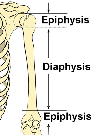
Of long bones, and epiphyseal ends of bones. Cells were isolated from the above figure 1. The diaphysis and the epiphysis (figure 6.3.1). The end of the long bone is the epiphysis and the shaft is the diaphysis. The blue represents additional matrix filling in the space btwn.
Bone Structure Anatomy Explained What Is Bone Marrow from www.teachpe.com The endosteum appears at the interface between the. It is found in bones such as the humerus and the. A long bone has diaphyseal bone is organized to create the best balance between weight and structural strength. Blood vessels and tissue surrounding the injured area bleed and if medullary lesions develop along the inner aspect of the cortical bones, especially in the long bones, endosteal scalloping may be observed. (see concentric and interstitial lamellae). Like the bone marrow, the periosteum and endosteum are enriched with mps to maintain skeleton homeostasis. Osteoclasts on the inside in the endosteum remove this bone to maintain the bone diameter. The diaphysis and the epiphysis (figure 6.3.1).
Long bones are formed from a cartilage model precursor by endochondral ossification (see the image below) and can range in size from a phalanx to a femur.
When osteoclasts start removing less bone, or osteoblasts start adding more bone, the. Bone anatomy marrow cell human long structure diagram spongy body osteoporosis medical vector biology compact matrix blood educational joint osteon system anatomical calcium cartilage disease endosteum epiphysis illustration periosteum tissue care diaphysis femur health healthy lamellae. Of long bones, and epiphyseal ends of bones. Make sure that you follow all the guidelines for biological drawings: They are one of five types of bones: Mesenchymal progenitors were isolated and identified. The endosteum can be seen in the t.s. This endosteal surface is usually resorbed during long periods of malnutrition, resulting in less cortical thickness. In an adult, most red blood cells are formed in the marrow in flat bones. 14 (makes up the whole area). Cells were isolated from the above figure 1. Periosteum and endosteum the external surface of bone is covered by the periosteum and its internal surface is lined by the endosteum. It is found in bones such as the humerus and the.
Structure of long bone although there are many different types of bones in the skeleton, we will endosteum: long bone diagram. □ the white, or yellow marrow fills up the medullary cavities.


0 Comments